
        
|
Please choose one of the following...
What is a virus..... The word "virus" is Latin for a poison or slimy liquid. A virus (plural: viruses- not viruses) is an infectious agent found in all life forms including humans, animals, plants and bacteria. It is a microbe which can harm any livng thing. Unlike bacteria, viruses cannot be killed by any known medicine. They are either made from DNA (deoxyribonucleuic acid) or RNA (ribonucleuic acid). This genetic material is protected by a protein called capsid. Sometimes, 100 viruses can fit into a bacteria. They cannot be seen by an ordinary microscope and can be seen by an electron microscope which shows false colours. The largest virus: poxvirus is 250 nanometres long and the smallest: poliovirus is 30 nanometres long. A million polioviruses can fit in this dot. Many microbiologists (scientists who study microbes) argue that viruses are not living things because they can only reproduce inside a cell. They have evolved to transmit their genes from one living thing to another to reproduce. Most of the time, a virus causes diseases by damaging cells. When they reproduce in a cell, they produce toxins which damage them. Some types of viruses can make cells reproduce uncontrollably and thus cause cancer. Chemists find it difficult to invent anti-viral medicines for 3 reasons: a) they are too stupid. b) it is difficult to kill a virus without killing a cell. c) some types of diseases are caused by viruses made out of different protective coatings. Lymphocytes (white blood cells which produce anti-bodies) can't kill some types of viruses because they change their protein coating every time e.g. common cold. The study of viruses has lead to important discoveries about the human body. What are they made out of? An individual virus is called a viruseson. A viruseson contain a gene, or to be more accurate, a genome in one of several forms. Unlike most cells which have a DNA form only, viruses can have DNA or RNA to record the genetic information used during reproduction. Most DNA viruses have double stranded DNA which can be linear or circular. Most RNA viruses have single stranded RNA. They are usually linear. RNA in virusesi can be segmented (with different genes in different molecules) or non-segmented (wit all the genes in the same single piece). The protective coat or capsid can be helical (spiral-shaped) or icosahedral (with 20 triangular sides). Capsids consist of one or different types of repeating units of proteins. The units are called protomers are capsomers while the proteins are called structural proteins. For some reason, virusesi have genes for the structure of the proteins and are not used; they are only found in infected cells. They are called non-structural proteins; they have information for the production of a viruseson. Some virusesons consist of nucleocapsids (DNA/RNA and capsids) only while others contain other structures. Some animal virusesi have a protective envelope needed to enter a cell's membrane (outer wall). In these envelopes, there are glycoproteins which is directed by the virus for the cell to make. These proteins combines a virus to its host cell.
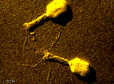
PICTURE:bacteriophages The most complicated virusesi are the bacteriophages, which reproduce inside bacteria. Incomplete viruses that cause diseases are called viroids and prions. Viroids are plant pathogens (microbes) that consist only of a circular, independently replicating RNA molecule. Replication The first contact between a virus particle and its host cell occurs when an outer viral structure docks with a specific molecule on the cell surface. For example, a glycoprotein called gp120 on the surface of the human immunodeficiency virus (HIV, the cause of acquired immune deficiency syndrome, or AIDS) viruseson specifically binds to the CD4 molecule found on certain human T lymphocytes (a type of white blood cell). Most cells that do not have surface CD4 molecules generally cannot be infected by HIV. Entering a cell After binding to an appropriate cell, a virus must cross the cell membrane (the outer part of a cell) . Some viruses accomplish this goal by fusing their lipid envelope to the cell membrane, thus releasing the nucleocapsid into the cytoplasm (the jelly-like substance found in a cell) of the cell. Other viruses must first be endocytosed (enveloped by a small section of the cell's plasma membrane that pokes into the cell and pinches off to form a bubblelike vesicle called an endosome) before they can cross the cell membrane. Conditions in the endosome allow many viruses to change the shape of one or more of their proteins. These changes allow the virus either to fuse with the endosomal membrane or to lyse the endosome (cause it to break apart), allowing the nucleocapsid to enter the cell cytoplasm. Division Once inside the cell, the virus replicates itself through a series of events. Viral genes direct the production of proteins by the host cellular machinery. The first viral proteins synthesized by some viruses are the enzymes required to copy the viral genome. Using a combination of viral and cellular components, the viral genome can be replicated thousands of times. Late in the replication cycle for many viruses, proteins that make up the capsid are synthesized. These proteins package the viral genetic material to make newly formed nucleocapsids. Escape PICTURE:HIV escaping from a white blood cell To complete the virus replication cycle, viruses must exit the cell. Some viruses bud out of the cell's plasma membrane by a process resembling reverse endocytosis. Other viruses cause the cell to lyse, thereby releasing newly formed virus particles ready to infect other cells. Still other viruses pass directly from one cell into a nearby cell. The virus replication cycle can be as short as a couple of hours for certain small viruses or as long as several days for some large viruses. Committing suicide Some viruses kill cells by causing severe damage resulting in cell lysis; other viruses cause the cell to kill itself in response to virus infection. This programmed cell suicide is thought to be a host defense mechanism to eliminate infected cells before the virus can complete its replication cycle and spread to other cells. Alternatively, cells may survive virus infection, and the virus can persist for the life of its host. Virtually all people harbor harmless viruses. Retroviruses, such as HIV, have RNA that is transcribed into DNA by the viral enzyme reverse transcriptase upon entry into the cell. (The ability of retroviruses to copy RNA into DNA earned them their name because this process is the reverse of the usual transfer of genetic information, from DNA to RNA.) The DNA form of the retrovirus genome is then integrated into the cellular DNA and is referred to as the provirus. The viral genome is replicated every time the host cell replicates its DNA and is thus passed on to daughter cells (cells produced after cell reproduction). Diseases
PICTURE: Chickenpox How do they enter the body Viruses can enter the body by several routes. Herpes simplex virus and poxviruses enter through the skin by touching an infected individual's skin. Ebola, hepatitis B, and HIV can be contracted from infected blood products. Hypodermic needles and animal and insect bites can transmit a variety of viruses through the skin. Viruses that infect through the respiratory tract are usually transmitted by airborne droplets of mucus or saliva from infected individuals who cough or sneeze. Viruses that enter through the respiratory tract include orthomyxovirus (influenza), rhinovirus and adenovirus (common cold), and varicella-zoster virus (chicken pox). Viruses such as rotavirus, coronavirus, poliovirus, hepatitis A, and some adenoviruses enter the host through the gastrointestinal tract. Sexually transmitted viruses, such as herpes simplex, HIV, and human papilloma viruses (HPV), gain entry through the genitourinary route. Other viruses, including some adenoviruses, echoviruses, Coxsackie viruses, and herpesviruses, can infect through the eye. Infection Virus infections can be either localized or systemic. The path of virus spread through the body in systemic infections differs among different viruses. Following replication at the initial site of entry, many viruses are spread to their target organs by the bloodstream or the nervous system. The particular cell type can influence the outcome of virus infection. For example, herpes simplex virus undergoes lytic replication in skin cells around the lips but can establish a latent or dormant state in nerve cells (located in ganglia) for extended periods of time. During latency, the viral genome doesn't do anything in the cell nucleus until a stimulus such as a sunburn causes a chemical reaction, leading to the lytic replication cycle. Once reactivated, the virus travels from the ganglia back down the nerve to cause a cold sore on the lip near the original site of infection. The herpesvirus genome does not integrate into the host cell genome. Acute and chronic Virus-induced illnesses can be either acute, in which the patient recovers promptly, or chronic, in which the virus remains with the host or the damage caused by the virus is irreparable. For most acute viruses, the time between infection and the onset of disease can vary from three days to three weeks. In contrast, onset of AIDS following infection with HIV takes an average of 7 to 11 years. Cancer Because a number of viruses have been shown to cause tumors in animal models, it is probable that many viruses have a key role in human cancers. AIDS
The human immunodeficiency virus (HIV), which causes acquired immune deficiency syndrome (AIDS), attacks CD4 T-lymphocytes, an important type of white blood cell. As a result, the bodys ability to resist opportunistic viral, bacterial, fungal, protozoal, and other infection is greatly weakened. Pneumocystis carinii pneumonia is the leading cause of death among people with HIV infection, but the incidence of certain types of cancers such as B-cell lymphomas and Kaposis sarcoma is also increased. Neurological complications and dramatic weight loss, or wasting, are characteristic of end-stage AIDS. HIV can be transmitted sexually; through contact with contaminated blood, tissue, or needles; and from mother to child during birth or breastfeeding. Not all HIV positive people necessarily have AIDS. At first humans may have fever for 2 weeks with a small number of CD4 T-lymphocytes (fewer than 200 per mm cubed). After that, a human will feel well for 7-20 years with a safe number of CD4 T-cells (200-500 per mm cubed). Soon, the number drops dramatically and a human dies from other diseases after a few weeks.
More than one species Some virusesalphaviruses and flaviviruses, for examplemust be able to infect more than one species to complete their life cycles. Eastern equine encephalomyelitis virus, an alphavirus, replicates in mosquitoes and is transmitted to wild birds when the mosquitoes feed. Thus, wild birds and perhaps mammals and reptiles serve as the virus reservoir, and mosquitoes serve as vectors essential to the virus life cycle by ensuring transmission of the virus from one host to another. Horses and people are accidental hosts when they are bitten by an infected mosquito, and they do not play an important role in virus transmission. Mad cow disease The most common human prion disease is Creutzfeldt-Jakob disease (CJD), which has a worldwide incidence of approximately one in a million individuals and is characterized by dementia. Scrapie is the most common prion disease in animals. Feed for cattle generated from scrapied sheep in Great Britain has resulted in the death of more than 150,000 cattle from bovine spongiform encephalopathy, or mad cow disease, since the discovery of the disease in 1986. It is not yet known if humans can develop CJD from consuming prion-contaminated beef, but several recent cases in Great Britain suggest this possibility. Defense Although viruses cannot be treated with antibiotics, which are effective only against bacteria, the body's immune system has many natural defenses against virus infections. Infected cells produce interferons and other cytokines (soluble components that are largely responsible for regulating the immune response), which warn nearby uninfected cells to mount their defenses, enabling uninfected cells to impair virus replication. Some cytokines can cause a fever in response to viral infection; increasing body temperature retards the growth of some types of viruses. B lymphocytes produce specific antibodies that can bind and inactivate viruses. Cytotoxic T cells recognize virus-infected cells and target them for destruction. However, many viruses have evolved ways to resist some of these host defense mechanisms. Medicines The development of antiviral therapies has been thwarted by the difficulty of generating drugs that can kill viruses only and leave cells alone. Therefore, most treatments for viral diseases simply cause relief from effects, such as fever, dehydration, and achiness. Nevertheless, antiviral drugs for influenza virus, herpesviruses, and HIV are available, and many others are in the experimental and developmental stages. Prevention Prevention has been a more effective method of controlling virus infections. Viruses that are transmitted by insects or rodent excretions can be controlled with pesticides. Successful vaccines are currently available for poliovirus, influenza, rabies, adenovirus, rubella, yellow fever, measles, mumps, and chicken pox. Vaccines are prepared from killed (inactivated) virus, live (weakened) virus, or isolated viral proteins (subunits). Each of these types of vaccines elicits an immune response while causing little or no disease, but there are advantages and disadvantages to each. Vaccination The principle of vaccination was discovered by British physician Edward Jenner. In 1796 Jenner observed that milkmaids in England who contracted the mild cowpox virus infection from their cows were protected from smallpox, a frequently fatal disease. In 1798 Jenner formally demonstrated that prior infection with cowpox virus protected those that he inoculated with smallpox virus (an experiment that would not be allowed because of its use of humans). In 1966 the World Health Organization (WHO) initiated a program to eradicate smallpox from the world. Because it was impossible to vaccinate the entire world population, the eradication plan was to identify cases of smallpox and then vaccinate all of the individuals in that vicinity. The last reported case of smallpox was in Somalia in October 1977. An important factor in the success of eradicating smallpox was that humans are the only host and there are no animal reservoirs for smallpox virus. The strain of poxvirus used for immunization against smallpox was called vaccinia. Introduction of the Salk (inactivated) and Sabin (live, attenuated) vaccines for poliovirus, developed in the 1950s by Jonas Salk and Albert Bruce Sabin, was responsible for a significant worldwide decline in paralytic poliomyelitis. However, polio has not been eradicated, partly because the virus can change their protein structure so that white blood cells won't be able to notice them and escape the host immune response. Influenza viruses mutate so rapidly that new vaccines are developed for distribution each year. Mutation Viruses undergo very high rates of mutation (genetic alteration) largely because they lack the repair systems that cells have to safeguard against mutations. A high mutation rate enables the virus to continually adapt to new intracellular environments and to escape from the host immune response. Discovery By the last half of the 19th century, scientists have discovered protozoa, fungi, and bacteria, all visible with a light microscope. In the 1840s, the German scientist Jacob Henle suggested that there were infectious agents too small to be seen with a light microscope, but for the lack of direct proof, his hypothesis was not accepted. Although the French scientist Louis Pasteur was working to develop a vaccine for rabies in the 1880s, he did not understand the concept of a virus. Key Discoveries During the last half of the 19th century, several key discoveries were made that set the stage for the discovery of viruses. Pasteur is usually credited for proving that organisms reproduce new organisms. The German scientist Robert Koch, a student of Jacob Henle, and the British surgeon Joseph Lister developed techniques for growing cultures of single organisms that allowed the assignment of specific bacteria to specific diseases. Wrong Conclusion The first experimental transmission of a viral infection was accomplished in about 1880 by the German scientist Adolf Mayer, when he demonstrated that extracts from infected tobacco leaves could transfer tobacco mosaic disease to a new plant, causing spots on the leaves. Because Mayer was unable to isolate a bacterium or fungus from the tobacco leaf extracts, he considered the idea that tobacco mosaic disease might be caused by a soluble agent, but he concluded incorrectly that a new type of bacteria was likely to be the cause. The Russian scientist Dimitri Ivanofsky extended Mayer's observation and reported in 1892 that the tobacco mosaic agent was small enough to pass through a porcelain filter known to block the passage of bacteria. He too failed to isolate bacteria or fungi from the filtered material. But Ivanofsky, like Mayer, was bound by the dogma of his times and concluded in 1903 that the filter might be defective or that the disease agent was a toxin rather than an organism. Controversy Unaware of Ivanofsky's results, the Dutch scientist Martinus Beijerinck, who collaborated with Mayer, repeated the filter experiment but extended this finding by demonstrating that the filtered material was not a toxin because it could grow and reproduce in the cells of the plant tissues. In his 1898 publication, Beijerinck referred to this new disease agent as a contagious living liquidcontagium vivum fluidcausing a 20-year controversy over whether viruses were liquids or particles. Proofs The conclusion that viruses are particles came from several important observations. In 1917 the French-Canadian scientist Félix H. d'Hérelle discovered that viruses of bacteria, which he named bacteriophage, could make holes in a culture of bacteria. Because each hole, or plaque, developed from a single bacteriophage, this experiment provided the first method for counting infectious viruses (the plaque assay). In 1935 the American biochemist Wendell Meredith Stanley crystallized tobacco mosaic virus to demonstrate that viruses had regular shapes, and in 1939 tobacco mosaic virus was first visualized using the electron microscope. |

 Most
viral infections cause no symptoms and do not result in disease. For example,
only a small percentage of individuals who become infected with Epstein-Barr
virus or western equine encephalomyelitis virus ever become ill. In contrast,
most people who are infected with measles, rabies, or influenza viruses develop
the disease. A wide variety of viral and host factors determine the outcome
of virus infections. A small genetic variation can produce a virus with
increased capacity to cause disease. Such a virus is said to have increased
virulence.
Most
viral infections cause no symptoms and do not result in disease. For example,
only a small percentage of individuals who become infected with Epstein-Barr
virus or western equine encephalomyelitis virus ever become ill. In contrast,
most people who are infected with measles, rabies, or influenza viruses develop
the disease. A wide variety of viral and host factors determine the outcome
of virus infections. A small genetic variation can produce a virus with
increased capacity to cause disease. Such a virus is said to have increased
virulence.